Inner banner

The brain’s white matter serves a vital purpose within the human body in that it transports impulses to gray matter cells. When a person suffers a periventricular leukomalacia injury, these functions are impaired. PVL is a strikingly common causal factor among children with Cerebral Palsy that leads to intellectual impairment and spasticity that require therapy and treatment.
Periventricular leukomalacia, or PVL, is a type of brain damage that involves the periventricular white matter of the brain. Damage to the white matter results in the death and decay of injured cells, leaving empty areas in the brain — called lateral ventricles, which fill with fluid (a condition called leukomalacia).
The brain primarily consists of white matter and gray matter. Gray matter has neural cell bodies, which can initiate nerve impulses, while white matter transports impulses between gray matter cells. The periventricular white matter that surrounds two horseshoe shaped cavities in the brain is primarily responsible for the transmission of nerve impulses that control motor function. Damage in this area can result in spasticity and intellectual impairment.
Myelin is an integral component of white matter that coats and essentially insulates cell pathways, promoting speedy transmission of nerve impulses. Damage to myelin slows and impedes nerve transmission, possibly impairing brain function.
Experts believe intrauterine infections are the underlying factor for periventricular leukomalacia. Membranes around the fetus are affected by the release of toxins, which travel through amniotic fluid to selectively injure areas of the developing brain. These toxins can also cause premature rupture of the membranes and premature birth.
Damage can occur at any time, but researchers have isolated stages of fetal development when the unborn child is particularly vulnerable to periventricular leukomalacia.
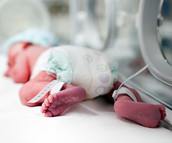
Experts report decreased blood flow or cell damage to periventricular tissue is an underlying cause of periventricular leukomalacia. Infants who are born before 32 weeks of gestation and are mechanically ventilated are at greatest risk for periventricular leukomalacia. Hypotension, hypoxemia, acidosis, and hypocarbia in ventilated premature infants can cause periventricular leukomalacia.
A number of other events also pose a significant increase in the likelihood of developing periventricular leukomalacia. Intrauterine infection, where abnormal bacteria can infect the amniotic fluid, is one factor; infection around the time of delivery also increases likelihood. This is more common during premature delivery. Other risk factors associated with periventricular leukomalacia include:
Periventricular leukomalacia can be difficult to detect in newborns, as very young infants typically do not show signs of impairment. Periventricular leukomalacia can resemble other conditions, and all cases are different. Intellectual impairment, developmental impairment, visual dysfunction, hearing impairment, and problematic coordination are common with periventricular leukomalacia.
Spastic diplegia (tight muscles and limbs that do not bend easily) is the most common type of Cerebral Palsy caused by periventricular leukomalacia; quadriplegia is the most severe.
Cranial ultrasounds, MRIs, and CT scans can identify periventricular leukomalacia. Because early ultrasounds may not reveal periventricular leukomalacia — it can take four to eight weeks for the condition to become detectable — infants with known risk factors are often tested approximately 30 days after birth. Medical histories and clinical exams can identify infants to be tested.
Babies diagnosed with periventricular leukomalacia, or at risk for periventricular leukomalacia, require special care after hospital discharge. Cranial ultrasounds performed too early may not detect periventricular leukomalacia. Frequent developmental assessments are performed if periventricular leukomalacia is suspected. Treatment is typically focused on managing symptoms through massage therapy, physical therapy, speech therapy, and treatment for visual dysfunction.
Intrauterine infection is difficult to detect, especially when it does not present noticeable symptoms. Although deemed impractical by some, others believe elaborate tests at five to seven months of pregnancy could detect and treat infection and potentially prevent many cases of periventricular leukomalacia. Work is underway to develop an effective method of screening and treating pregnant women at risk for intrauterine infection.
Because the terminology used is so specific, yet remarkably similar, terms such as brain defect, brain malformation and brain lesion can seem confusing. It is helpful to know the difference between the terms when attempting to understand the cause of cerebral palsy.
Brain development begins shortly after conception. A relatively small number of cells divide and multiply into billions of cells. A small strip of tissue rolls into a neural tube. One end develops into the brain, the other into the spinal cord. Throughout, different types of cells form, group, and migrate to form various regions of the brain. The brain is considered fully developed two to five years after birth.
Brain defects are irregularities in the brain structure that typically cause impairment. Defects can occur from malformation, injury, illness or disease. The degree of impairment often is linked to the severity of damage. A brain sometimes compensates for defects, in essence, by “rewiring” to bypass or compensate for damaged areas. For this reason, beginning treatment as early as possible is typically recommended.
Brain malformations are defects that occur through abnormal development of the brain. Although defects can occur anytime during fetal development, in the first 20 weeks the infant is most vulnerable; any malformation that occurs while the neural tube is forming can have permanent consequences. Brain malformations may result in undeveloped areas, abnormal growth, malformation, or improper brain division into hemispheres and lobes.
Brain lesions are defects that occur from injury or disease. The cause of brain lesions during fetal development include bleeding in the brain, infections, toxins, asphyxia, and many others. Lesions typically result from an incident or event that causes brain tissue to die. Holes, which often fill with liquid, are left behind to form cysts.
Cerebral Palsy is caused by brain injury or brain malformation that occurs before, during, or immediately after birth while the infant’s brain is under development. But how a brain injury affects a child’s motor functioning and intellectual abilities is highly dependent on the nature of a brain injury, where the damage occurs, and how severe it is.
Cause
Damage to the white matter tissue in the brain
Periventricular Leukomalacia
Brain hemorrhage
Intraventricular Hemorrhage
Lack of oxygen to the brain, asphyxia, or intrapartum asphyxia
Hypoxic-Ischemic Encephalopathy
Brain malformation or abnormal brain development.
Cerebral Dysgenesis

Cerebral Palsy risk factors are events, substances or circumstances that increase the chances of a child developing Cerebral Palsy. They can be avoidable, or unavoidable. A risk factor does not ensure a child will develop Cerebral Palsy; it means chances are higher than if that risk factor was not present. Likewise, the absence of risk factors does not ensure that a child will not develop Cerebral Palsy. Have you been exposed to the following risk factors?
A risk factor does not ensure a child will develop Cerebral Palsy; it means chances are higher than if that risk factor was not present. Likewise, the absence of risk factors does not ensure that a child will not develop Cerebral Palsy.
Any exposure to risk factors prior to conception and during pregnancy should be immediately discussed with a doctor in order to treat and minimize risk. The Cerebral Palsy Risk Factor Checklist helps parents determine if they may have been exposed to risk factors for Cerebral Palsy.